Transmission electron microscopy (TEM) holds the potential to resolve many questions in life science by providing a close view of the molecular machinery of the cell. Although biological macromolecules are almost invisible in TEM, their structure is imprinted in the transmitted electron beam as small variations in the phase of the electron wave function.
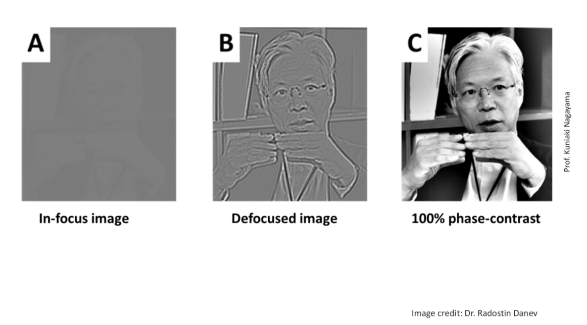
Like any other wave, electrons interfere with each other constructively or destructively depending on their relative phases. Since biological macromolecules depart only a very small phase shift to electrons in TEM, it is nearly impossible to see contrast in the final image, given by the complex square of the superposition of electron wave functions.

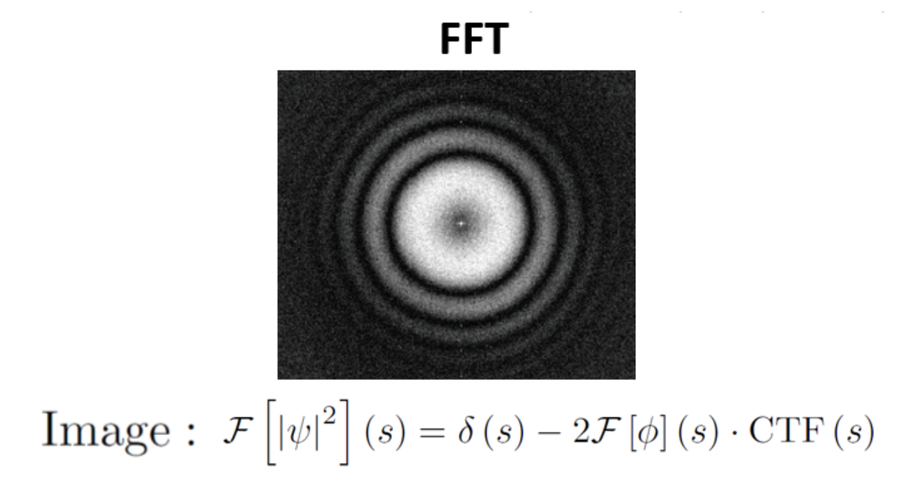
One way that researchers have sought to mitigate this issue is by using defocus-based phase contrast. That is, by setting the electron microscope slightly out of focus, the resulting contrast transfer function alters the Fourier transform of the final image so that there is good contrast for some spatial frequencies (white).
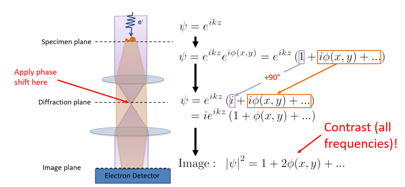
Another way to obtain phase contrast is with the use of a phase plate, which is placed in the diffraction plane of the electron microscope and thus imparts an additional phase shift to only the part of the beam that is unscattered by the specimen, shown in purple below. For ideal phase contrast, this additional phase shift is 90 degrees.
Several phase plate designs for electron microscopy have been attempted in the past, but their performance has been limited due to the demanding environment of the electron microscope. Electrons cause damage and charging of physical objects and are highly sensitive to spurious magnetic fields. Our group is pursing an alternative phase plate design which circumvents these issues by using an intense, tightly-focused laser beam to provide phase contrast. You can read more about the laser phase plate here.